 News
News
News
News
News
News
 News
News
Congratulations Nya!
 News
News
Congratulations Dr. Jahed!
 News
News
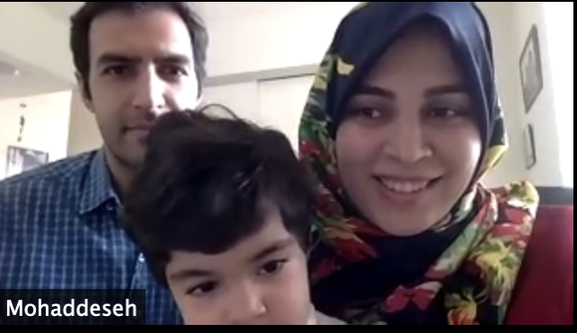
Congratulations Dr. Peyro!